Award winning paper uses equine standing MRI for diagnostic imaging above the foot

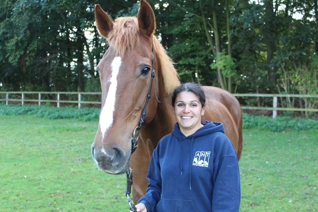
At Europe’s largest Equine Veterinary Conference – the British Equine Veterinary Association (BEVA) Congress held in September – Carolyne Tranquille of the Animal Health Trust was presented with the Sam Hignett Award for the best clinical research presentation. The paper was the result of case reviews of pain in the proximal metacarpal area in horses and used standing MRI to identify the lesions that were present. It confirmed how standing MRI is being used to produce diagnostic images above the foot, at the level of the knee.
Hallmarq Veterinary Imaging launched its standing equine MRI in 2001 and it has revolutionised the veterinary approach to equine lameness, allowing diagnostic quality images to be produced without anaesthesia. Many practices tend to focus on the benefits of scanning the foot area – which accounts for around 50% of all lameness cases in horses – but this study shows how imaging of the standing sedated horse can also yield diagnostic information on structural abnormalities as high as the knee.
The team behind the paper was led by Rachel Murray, Associate of European College of Veterinary Diagnostic Imaging and RCVS Specialist in Equine Orthopaedics and includes Carolyne Tranquille, Vicki Walker, Jack Tacey, all of the Animal Health Trust, Rebecca Milmine of Dubai Equine Hospital, Dubai and Nick Bolas of Hallmarq Veterinary Imaging.
Proximal metacarpal problems are a common cause of lameness, particularly in sporting horses and it can be difficult to identify lesions using other diagnostic techniques or imaging modalities. The paper was able to use standing MRI scans to show that abnormalities of the third metacarpal (MC3) were most frequently detected in the palmaro-medial quarter. In over half of all the cases reviewed (53%) there were abnormalities in both MC3 and the suspensory ligament.
Carolyne said the team were honoured to win the award, "It is exciting that the advances in technology have meant that it is possible to image the proximal metacarpal region using MRI in enough standing horses that we can do studies like this one, which improve our understanding of the types of pathology occurring in this region."
By using MRI for these types of lesions vets will be able to define the precise structural abnormalities present and give an accurate prognosis, helping horse owners make decisions about their horses’ future capabilities. To find out more about standing equine MRI and Hallmarq Veterinary Imaging visit www.hallmarq.net or call +44 1483 877812.
Tags:


